Original Article


Frequency of Neurogenic Strabismus in Al-Ibrahim Eye Hospital, Karachi


Shua Azam, Priyanka, Muhammad Qasim


Pak J Ophthalmol 2019, Vol. 35, No. 2

 . . . . . . . . . . . . . . . . . . . . . . . . . . . . . . . . . . . . . . . . . . . . . . . . . . . . . . . . . . . . .. . .. . . . . . . . . . . . . . . . . . . . . . . . . . . . . . . . . . .  . .

See end of article for

authors affiliations


…..………………………..


Correspondence to:

Shua Azam

M. Phil Optometry

Senior Lecturer

ISRA School of optometry,

Al-Ibrahim Eye Hospital, Karachi

Email: optomshuaazam@gmail.com


Purpose: To determine the frequency of Neurogenic Strabismus presenting at the Orthoptics Clinic in Al-Ibrahim Eye Hospital, Malir Karachi.

Study Design: Cross-sectional study.

Sampling Technique: Non-probability convenient sampling.

Place and Duration of Study: Orthoptics Clinic of Al-Ibrahim Eye Hospital (AIEH) Karachi, Pakistan from May to October, 2018.

Material and Methods: This study included 349 subjects age ranged from 5 to 75 years. All ocular examinations for strabismus were performed, including cover test, prism cover test and Hess chart. Demographic features and etiologies were recorded, and the causes of extra ocular muscle palsies were grouped as; trauma, diabetes, hypertension and others. SPSS version 20.0 was used to analyze the data.

Results: Frequency of neurogenic strabismus was found to be 6%. Out of 21 subjects, 8 (38.1%) subjects had diabetes, followed by 2 (9.5%) subjects with hypertension, 5 (23.8%) subjects with ocular trauma and 6 (28.6%) subjects with other causes. The most commonly affected side was the right eye seen in 13 (61.9%) subjects. The most common ocular motor nerve involved was abducent (sixth) nerve in 13 (61.9%) subjects, followed by oculomotor (third) nerve in 4 (19%) subjects. Out of the patients with third nerve palsy 3 (14.3%) subjects had pupil sparing and  only 1 (4.8%) subject had no  pupil sparing.

Conclusion: Sixth nerve was the most common nerve involved and most common etiology was uncontrolled diabetes.

Keywords: Neurogenic Strabismus, Cranial nerve palsies, Paralytic strabismus.


Strabismusis is a very common ocular cause of visual impairment in optometry and ophthalmology. The prevalence of strabismus worldwide is reported as 5.7%1. Strabismus or squint is a disorder in which the eyes are not properly aligned with each other. It involves a lack of coordination between muscle movements of two eyes. It can be due to either an imbalance of muscles or disruption in the nerve supply2. Paralytic or incomitant strabismus occurs when there is limitation of ocular movement. Palsy disrupts the maintenance of binocular single vision and due to loss of fusional amplitude resulting in diplopia (double vision) which may be compensated by abnormal head posture3. Many different treatment options are available to resolve the issue, including occlusion, refractive correction, prisms, vision therapy and surgical intervention4.

A paralytic deviation undergoes several stages. The first stage is characterized by limitation of movement affecting one muscle, as a rule and secondly by over action of contralateral synergist. During this stage the law of equal innervation exhibits. The third stage is contracture of ipsilateral antagonist that shows the reciprocal innervation to the muscle. And lastly, secondary inhibition of contralateral antagonist occurs because the contracted antagonist in affected eye requires less innervation. All these stages result in an angle of deviation which increases on movement of the eyes in the direction of limitation and decreases when they move away from the affected side. Moreover secondary deviation is seen which is always greater than primary deviation. Secondary deviation is assessed by fixing the affected eye while primary deviation is assessed by fixing the normal eye4.

The paralysis of abducent nerve can be either congenital or acquired5 and the most common cause of 4th nerve palsy is congenital6. Common causes for pupil-sparing pathologies are diabetic neuropathy, myasthenia gravis, atherosclerosis, chronic progressive ophthalmoplegia and vasculopathies. On the other hand, the most common causes of non-pupil sparing oculomotor palsy are tumor, followed by vascular lesions (posterior communicating aneurysms, and then distal basilar artery aneurysms)7.

The rationale of the study was to collect data about cases with neurogenic strabismus so that we can manage them better. This study was carried out to identify patients in the orthoptic clinic who had ocular motility problems due to the neurogenic causes.


MATERIAL AND METHODS

It was hospital-based, cross-sectional study conducted at Orthoptics Clinic of Al-Ibrahim Eye Hospital (AIEH) Karachi, Pakistan by using non-probability, convenient sampling technique from May to October 2018. Ethical approval was given by Research Ethical Committee (REC) of Isra Postgraduate Institute of Ophthalmology. 349 patients who visited orthoptics clinic during period of data collection were included. The inclusion criteria were subjects between 5-75 years of age who had manifest neurogenic strabismus, no history of previous squint surgery or other ocular disease. Subjects with history of trauma, diabetes and hypertension were included as well. The exclusion criteria included subjects with latent and puesdo strabismus and syndromes.

All the subjects were examined after obtaining fully informed written consent. The protocol for examination for all patients included the demographic data, history of onset, type of squint. All this was retrieved from the case notes. History revealed whether the patient had trauma, diabetes or hypertension. Visual acuity of every patient was checked and recorded separately both for near and distance, with and without glasses. Then orthoptic assessment was done to evaluate the type of palsy which included cover/uncover test, ocular motility, prism cover test and pupillary reflex test. Cover test was assessed to check the eye affected in primary position, angle and type of tropia with occluder and fixation targets both in distance and near. Extra-ocular motility test in all gazes was checked first in versions to check any limitation (underactions) with secondary angle of deviation (overactions) and then ductions were checked by occluding one eye to confirm the limitation of gaze. Hess chart was performed to make the final diagnosis by correlating all the tests results. The anterior segment was also examined with a slit-lamp by an ophthalmologist to exclude any ocular disease and the refraction (dry or cycloplegic) was also assessed by optometrist. Data analysis was done on statistical package for social sciences (SPSS) version 20.0. All continuous variables were presented as mean ± standard deviation. The entire categorical variables were shown as frequency and percentages. Statistical charts were presented in the form of Bar chart & Pie chart.


RESULTS

A total of 21 subjects among 349 subjects fulfilled the inclusion criteria for the study. Among them, 5 were females, and 16 were males. The mean age of onset was 35.3 years, ranging between 5-75 years. The frequency of neurogenic strabismus was found to be 6%. Out of 21 subjects, 8 (38.1%) subjects were found to have diabetes, 2 (9.5%) subjects had hypertension, 5 (23.8%) subjects had ocular trauma and 6 (28.6%) subjects had other causes as shown in Figure 1. The most affected eye was right eye in 13 (61.9%) subjects as shown in Figure 2. At the end of the examination the most commonly seen manifest deviation on cover test was esotropia in 13 (61.9%) subjects, followed by exotropia in 4 (19%), hypotropia in 2 (9.5%), hypertropia in 1 (4.8%) and combined in 1 (4.8%) subject as shown in Figure 3. The diagnosis of all subjects on Hess chart and other tests showed the most common ocular motor nerve involved was abducent (sixth) nerve in 13 (61.9%) subjects, followed by oculomotor (third) nerve in 4 (19%) subjects. Out of the patients who had third nerve palsy 3 (14.3%) subjects had pupil sparing while only 1 (4.8%) subject had no pupil sparing. Double elevator palsy was seen in 2 (9.5%) subjects, there was a single case (4.8%) of fourth nerve palsy and combined nerve involvement was seen in 1 (4.8%) subject as shown in Figure 4.


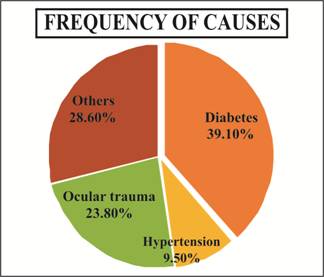
Fig. 1: Frequency of Causes of Palsies.


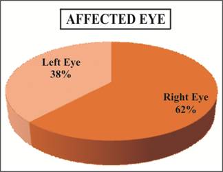
Fig. 2: Distribution of Affected Eye.


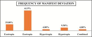
Fig. 3:   Frequency of Manifest Type of Deviation on Cover Test.


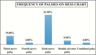
Fig. 4:   Frequency of Distribution of Palsies tested on Hess Chart.


Table 1: Palsy versus gender cross tabulation.


Palsy  Versus Gender Cross tabulation

Type of Palsy

Gender

Total

Male

Female

Third Nerve Palsy With no Pupil involvement

  1

0

  1

Third Nerve Palsy With Pupil involvement

  3

0

  3

Fourth Nerve Palsy

  1

0

  1

Sixth Nerve Palsy

10

3

13

Double Elevator Palsy

  1

1

  2

Combined

  0

1

  1

Total

16

5

21


DISCUSSION

Many studies are available which were conducted in different clinics of the world and show variety of results. According to a survey of 2007, the higher prevalence of paralytic strabismus was found to be 10%8 and a study in Turkey showed low prevalence about 4.75%9. However, in this study the frequency of neurogenic strabismus was found to be 6% as compared to the study that done in Gaza which constitutes 7.8%10. As such the differences in strabismus frequencies are due to changes in parameters used for diagnosis, ethnicity and time duration.

In this study, the frequency of neurogenic strabismus was more common in males. In males, it was 72.2% and in females 23.8%, which correlates with studies conducted in Lahore that showed the same ratio of males (74.3%) and females (25.7%)11,12. In the present study, right eye was more affected in 62%, while left eye in 38% and there was no case of bilateral involvement. While in previous studies left eye was commonly affected as compared to right eye9,13.

In this study, the most common cause was found to be diabetes. Previous studies showed the common cause of etiology was vascular diseases which includes hypertension and diabetes both14-17. Whereas, trauma was also common cause in many studies8,18,19.

A similar study in India showed the most common nerve involved was 6th nerve in 46.7%, followed by 3rd nerve 23.3%, combined nerve involvement 20% and 4th nerve 10%8,14. A study in Korea, showed the third nerve was commonly affected nerve among all12. Another study in Korea, showed the equally affected ratio of 6th and 4th nerve palsies13. Many studies giving the prevalence of third, fourth, and sixth nerve palsies had higher incidence of sixth nerve palsy followed by third and then fourth nerve palsies11,20. In some studies fourth nerve was more prevalent6,21. A study was conducted in China, which included all patients who had head trauma that showed the highest incidence of paralysis of third nerve (54.8%) followed by fourth nerve (45.2%) of all cases22.


CONCLUSION

The sixth nerve was the most commonly involved nerve in our patients. The most common etiology was uncontrolled diabetes.


Conflict of Interest

None.

ACKNOWLEDGEMENTS

Special thanks to

Professor Dr. Mohammad Saleh Memon

Dr. Abdul Hameed Talpur


Author’s Affiliation

Shua Azam

MPhil optometry

Senior lecturer

ISRA School of optometry,

Al-Ibrahim eye hospital Karachi


Priyanka

BS vision sciences

Optometrist intern

Al-Ibrahim eye hospital Karachi


Muhammad Qasim

MPH. BS vision Sciences

Assistant professor

ISRA School of optometry,

Al-Ibrahim eye hospital Karachi


Author’s Contribution

Shua Azam

Study design, article review, manuscript writing.


Priyanka

Review of literature

Data collection.


Muhammad Qasim

Data analysis and critical review.


REFERENCES

1.        Khorrami-Nejad M, Akbari MR, Khosravi B. The Prevalence of Strabismus Types in Strabismic Iranian Patients. Clinical Optometry, 2018; 10 (1): 19-24.

2.        Behera S, Bijaya KD, Chowdhury RK, Sar M. A Clinico-Anatomical Study of Strabismus in a Tertiary Care Hospital. Journal of Dental and Medical Sciences, 2014; 13 (1): 32-35.

3.        Anson AM, Davis H. Diagnosis and Management of Ocular Motility Disorders, 3rd ed. Blackwell Science Ltd: UK; 2001.

4.        Dasinger KD. Intermittent Exotropia: Management Options and Surgical Outcomes. J of Beh Opt. 2012; 23 (2): 44.

5.        Kasturi N. Congenital sixth nerve palsy with associated anomalies. Indian J Ophthalmol. 2017; 65 (1): 1056-7.

6.        Sekeroglu HT, FEBO, Turan KE, Arslan U et al. Etiology of Fourth and Sixth Nerve Palsies: a Single Ophthalmology Clinic’s Perspective. International Journal of Ophthalmology and Clinical Research 2014; 1 (1): 1-3.

7.        Lai G, Rodriguez MI, Scumpia AJ. Oculomotor Nerve Palsy Secondary to Cavernous Internal Carotid Aneurysm. Clin Pract Cases Emerg Med. 2018 Jan 9;2(1):93-94.

8.         Stidwill D. Epidemiology of Strabismus. Opthalmic and Physiological Optics, 2007; 17 (6): 536-539.

9.        Niyaz L, Gul A and Ariturk N. Frequency and Etiology of Paralytic Strabismus. Austin Journal of Clinical Ophthalmology, 2015; 2 (1): 1-2.

10.     Abuimara A. Relative prevalence of various types of strabismus in patients attending NGO's medical centers in Gaza Strip. Science Journal of Public Health, 2015; 3 (1): 1-5.

11.     Valsa TS, Susan P, Sreelatha KC. Clinical profile of third, fourth, and sixth cranial nerve palsies presenting to a tertiary care ophthalmic center. International Journal of Scientific Study, 2017; 5 (3): 93-97.

12.     Suman A, Nabin P, Gauri SS, Ananda KS. Clinical Profile of Extraocular Muscle Palsy: A Retrospective Study. Optometry & Visual Performance, 2013; 1 (6): 198-201.

13.     Taju S, Kebede W. Causes of cranial nerves III, IV and VI paralysis among Ethiopian patients presented at Menelik II hospital. Ethiopian Medical Journal, 2017; 56 (1): 23-29.

14.     Ho TH, Lin HS, Lin MC, Sheu SJ. Acquired paralytic strabismus in Southern Taiwan. J Chin Med Assoc. 2013 Jun; 76(6): 340-3.

15.     Kiyoung K, Sung RN. Clinical Course and Prognostic Factors of Acquired Third, Fourth, and Sixth Cranial Nerve Palsy in Korean Patients. Korean Journal Ophthalmology, 2018; 1 (1): 1-7.

16.     Ji SJ, Dae HK. Risk Factors and Prognosis of Isolated Ischemic Third, Fourth, or Sixth Cranial Nerve Palsies in the Korean Population. Journal of Neuro-Ophthalmology, 2015; 35 (1): 37-40.

17.     Kumar KH, Bhanu KBC, Ashok R. Clinical Study of 3rd, 4th and 6th Cranial nerve Palsies Leading to Visual Disturbances. International Journal of Contemporary Medical Research 2018; 5 (4): 10-12.

18.     Anayat A, Sadiq MAA. Ocular Motility Problems after Head Trauma. Ophthalmology Pakistan, 2015; 5 (3): 50-53.

19.     Park UC, Kim SJ, Hwang JM, Yu YS. Clinical features and natural history of acquired third, fourth, and sixth cranial nerve palsy. Eye (Lon) 2008; 22 (5): 691-696.

20.     Rowe F. Prevalence of Ocular Motor Cranial Nerve Palsy And Associations Following Stroke. Eye, 2011; 25 (1): 881-887.

21.     Bolutine O, Tinly C, Grotte R. Paralytic Strabismus in South African Black and Mixed Race Children – A 15-year Clinic-based Review. Ophthalmic Epidemiology, 2012; 19 (6): 396-400.

22.     Guichen Li, Xiaobo Z, Sun Y, Gao X, Zhang Y, Houn K. Ocular Movement Nerve Palsy After Mild Head Trauma. World Neurology, 2016; 94 (1): 296-302.